Global·EN
CN EN
NEOSCAN 台式显微CT 22
NEOSCAN desktop high-resolution Micro CT
NEOSCAN Desktop High-resolution Micro CT

NEOSCAN Micro CT technology combines X-ray imaging and computerized reconstruction to provide high-resolution 3D imaging and analysis of objects in a non-destructive manner.

What can micro CT do

High-resolution, non-destructive acquisition of the following information about the sample:

  • 01. Structural Information
    • Diameter
    • Volume
    • Surface area
    • Roundness
    • Connectivity
    • Spatial distribution
  • 02. Density Information
    • Cavity Pore
    • Elemental weight
    • Spatial distribution
  • 03. Three-dimensional modeling
    • Finite element analysis
    • 3D Printing
Product Presentation
Product Lists
Product Advantages
Product Advantages
Application Fields
Application Fields

NEOSCAN Micro CT can obtain structural information (cavities and pores) and density information (compositional differences) without destroying the sample.
At the same time, a 3D model can be output for simulation and analysis, which has a wide range of applications in many industries:
Application Cases
Application Cases
  • Foodstuffs
    Ice Cream
  • Composite Material
    Carbon Fiber
  • Composite Material
    Glass Fiber
  • Biotechnology
    Beetles
  • Geology
    Reservoir
  • Geology
    Shale
  • Medical
    Medicine Pill
  • Dental
    Toothless
Foodstuffs
Ice Cream
Composite Material
Carbon Fiber
Composite Material
Glass Fiber
Biotechnology
Beetles
Geology
Reservoir
Geology
Shale
Medical
Medicine Pill
Dental
Toothless
Principles of Operation
How Micro CT works
  • 01
    Scan

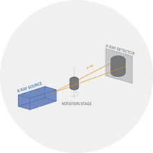
    During the scanning process, the sample stage is rotated in fixed steps and angles. After each rotation, the X-ray beam passes through the sample and the detector records the corresponding projection data. With multiple rotation angles and projection data collection, information about the projection of the sample in different directions can be obtained.
  • 02
    Reconstruction

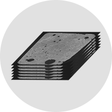
    The collected projection data is used to reconstruct the image using a computerized reconstruction algorithm. The most commonly used algorithm is Filtered Back Projection, which reconstructs a 3D image of the sample by filtering and back-projecting the projection data.

  • 03
    Image Processing and Analysis

    The reconstructed 3D image can be subsequently processed through image processing and analysis, including denoising, enhancement, segmentation, 3D visualization and other operations, in order to better observe and analyze the internal structure and characteristics of the sample.

01
Scan

During the scanning process, the sample stage is rotated in fixed steps and angles. After each rotation, the X-ray beam passes through the sample and the detector records the corresponding projection data. With multiple rotation angles and projection data collection, information about the projection of the sample in different directions can be obtained.

02
Reconstruction

The collected projection data is used to reconstruct the image using a computerized reconstruction algorithm. The most commonly used algorithm is Filtered Back Projection, which reconstructs a 3D image of the sample by filtering and back-projecting the projection data.

03
Image Processing and Analysis

The reconstructed 3D image can be subsequently processed through image processing and analysis, including denoising, enhancement, segmentation, 3D visualization and other operations, in order to better observe and analyze the internal structure and characteristics of the sample.

Online Message
Online Message

If you want to know more about the product information, book a sample or consult the after-sales service, you are welcome to call our hotline 400 857 8882 or leave us a message online, we receive your message will be the first time to reply to you.

  • *
  • *
  • *
  • *